+34 620 10 75 37info@nanbiosis.com

News U8

Microfluidic device that reproduces the blood-retinal barrier

The use of In vitro testing with living cells as an alternative to animal research has limitations like the difficulty to reproduce the interaction of cells. To overcome it, scientists are working on the development of systems that simulate and reproduce functions of tissues and organs in conditions very similar to reality. They are called organ-on-a-chip, which include microenvironments and microarchitectures that simulate the state of tissues and living organs.

Scientists of NANBIOSIS Unit 8 have published in an article, cover of the magazine “Lab on a Chip”, the “proof of concept” of a microfluidic device that reproduces the blood-retinal barrier, that is, a microchip that allows us to reproduce what happens ” in vivo ‘in the retina. This device can be an essential tool that revolutionizes experimentation ‘in vitro’.

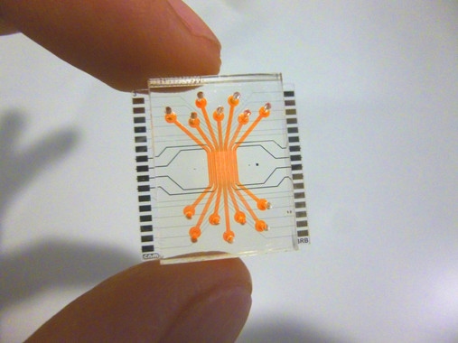
José Yeste, researcher of the CIBER-BBN, explains that the micro device consists of several parallel compartments, in which different types of cells have been cultivated to emulate the structure of cellular layers of the retina. They are endothelial cells, that is, they form the internal part of the barrier, in contact with the blood capillaries, through which oxygen and nutrients reach the retina. In addition, it is also composed of neuronal cells (which form the neuroretina), and pigment epithelial cells, which constitute the outer layer. The compartments are interconnected in their lower part by a network of micro-grooves, so as to allow an intercellular communication through the exchange of signalling molecules between cells. Thus, cells can send their signals to others and interact, much like they would in a living organism. In addition, the micro device allows the endothelial cells to be subjected to the mechanical stimulus induced by the flow to emulate a more physiological microenvironment.

“Within the body, the endothelial cells that line the inside of blood vessels are subject to the mechanical stimulation of blood circulation. In cell cultures that do not reproduce this flow, the cells are as ‘lethargic’, and do not respond in the same way they would in real conditions, “explains Rosa Villa, Scientific Director of NANBIOSIS Unit 8 and leader of the group of Biomedical Applications of the Microelectronics Institute of Barcelona of the CSIC.

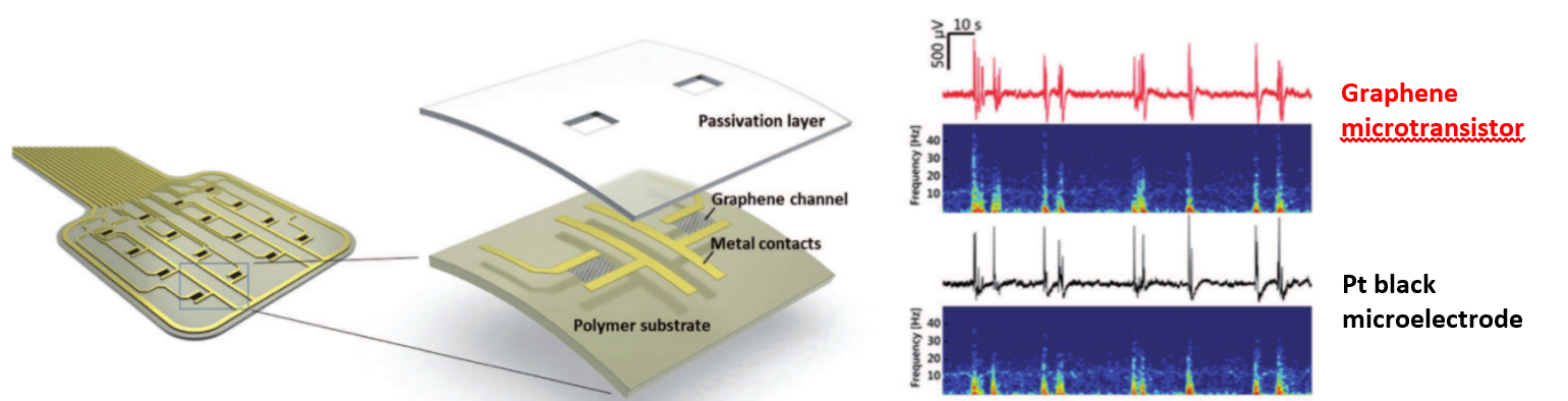
Scientists have evaluated the correct formation of the blood-retinal barrier by performing permeability, electrical resistance tests, as well as protein expression of tight junctions between cells. These tests were intended to verify that the barrier is well formed, that it has closed but maintains the natural permeability, sufficient to allow the passage of nutrients and oxygen, and that the cells are in contact and interact with each other.

This work has been developed in the ICTS NANBIOSIS, more specifically in Unit 8 of Micro-Nano Technology located in the IMB-CNM. It is also part of the results of the CIBER intramural project called Micro BRB: Microfluidic model of retinal neurovascular unit to identify new therapeutic targets in diabetic retinopathy (2016-2017) in wich also  participates Unit 3 of NANBIOSIS

Source: http://noticiasdelaciencia.com/not/27155/un-microchip-microfluidico-reproduce-la-barrera-de-la-retina-humana/

Article of reference:

A compartmentalized microfluidic chip with crisscross microgrooves and electrophysiological electrodes for modeling the blood–retinal barrier. Jose Yeste, Marta arcía-Ramírez, Xavi Illa, Anton Guimerà, Cristina Hernández, Rafael Simó and Rosa Villa. DOI: 10.1039/C7LC00795GLab Chip, 2018, 18, 95-105

Read More

Graphene transistors as eficient transducers for electrocorticography

Researchers of Micro-Nano Technologies Unit (U8) of NANBIOSIS, are co-authors of a new paper published in Advanced Functional Materials. Neuroelectronic interfaces bridge the central nervous system to the outside world and hold great potential for functional restoration in persons with paralysis, other forms of motor dysfunction, or limb loss. Neuroscientists and neurosurgeons are thus looking for technologies that could ideally record the whole brain with a high spatial and temporal resolution. Electrocorticography (ECoG), the practice of placing arrays of large-diameter electrodes (few millimeters) directly on the cortex is the current clinical solution to obtaining brain recordings with high temporal resolution.

Recent research from the CIBER-BBN and  IMB-CNM Biomedical Applications group (IP Rosa Villa)  coordinator of NANBIOSIS Unit 8, in collaboration with ICN2 (IP JA Garrido) , IDIBAPS  (IP MV Sanchez Vives) and INSERM (IP B Yvert) groups,  has focused on the development of graphene technology for electrocorticography. Specifically, flexible graphene transistor arrays have been fabricated and applied to the in vivo measurement of local field potentials.

Graphene is one of the most promising material candidates for neural interfacing thanks to its biocompatibility, low dimensionality and mechanical properties. Additionally, graphene exhibits extraordinary electrical properties such as high carrier mobility and chemical stability, features that only few materials can offer therefore helping to create a very intimate interface between the tissue and the transducing system.

However, previous in vivo studies using single layer CVD graphene have used an electrode configuration. Instead, here they propose the use of a transistor configuration. The main reason for this choice is certainly the local preamplification inherent to a transistor. As a consequence, less environmental noise is picked by the device.

Their work presents a complete description of the fabrication technology, the operation of graphene solution-gated field-effect transistors (SGFET) in saline solution and of the custom characterization electronic system. The devices are finally used in in vivo experiments in which the transconductance and noise are first characterized during slow-wave activity followed by the recording of visual and auditory evoked activity as well as of synchronous activity in a rat model of epilepsy. An in-depth comparison of the signal-to-noise ratio of graphene SGFETs with that of platinum black electrodes confirms that graphene SGFET technology is approaching the performance of state-of-the art neural technologies.

Full details of the fabrication, characterization and in vivo performance of the flexible graphene transistor probes can be found in the paper below.

Hébert, C., et al., Flexible Graphene Solution‐Gated Field‐Effect Transistors: Efficient Transducers for Micro‐Electrocorticography. Advanced Functional Materials, 2017.

Read More

Inkjet-Printed Sulfide-Selective Electrode

Gemma Gabriel, Scientific Coordinator of Unit 8 of NANBIOSIS is coauthor of the article  “Inkjet-Printed Sulfide-Selective Electrode” recently publish by ACS Publications in Analitical Chemistry.  Anal. Chem.201789 (22), pp 12231–12236  

DOI: 10.1021/acs.analchem.7b03041


Read More

Posters presentation by NANBIOSIS Units in CIBER-BBN ANNUAL CONFERENCE 2017

Last 13 and 14 of November, CIBER-BBN  has celebrated its 11th Annual Conference in Hotel Santemar in Santander. In this conference there was a poster session with the participation of the following Units of NANBIOSIS. Special mention deserves Unit 1 with Neus Ferrer as Director and  Paolo Saccardo as Coordinator (in the picture):

Posters:

U1. Protein Production Platform (PPP):

Engineering protein complexes as nano- or micro-structured vehicles or drugs for human and veterinary medicine. Ugutz Unzueta, Naroa Serna, Laura Sánchez-García, José Vicente Carratalá, Olivia Cano-Garrido, Mercedes Márquez, Paolo Saccardo, Rosa Mendoza, Raquel Díaz, Héctor, López-Laguna, Julieta Sánchez, Anna Obando, Amanda Muñoz, Andrés Cisneros, Eric Voltà, Aida Carreño, José Luis Corchero, Neus Ferrer-Miralles, Esther Vázquez, Antonio Villaverde.

Units  U1. Protein Production Platform (PPP) and U18. Nanotoxicology Unit:

Intrinsic functional and architectonic heterogeneity of tumor-targeted protein nanoparticles. Mireia Pesarrodona, Eva Crosa, Rafael Cubarsi, Alejandro Sanchez-Chardi, Paolo Saccardo, Ugutz Unzueta, Fabian Rueda, Laura Sanchez-Garcia, Naroa Serna, Ramón Mangues, Neus Ferrer Miralles, Esther Vázquez, Antonio Villaverde.

Units U3. Synthesis of Peptides UnitU6. Biomaterial Processing and Nanostructuring Unit, and U20. In Vivo Experimental Platform:

Synthesis of different length monodisperse COL-PEG-PEPTIDE to increase biodisponibility of multifunctional nanovesicles for Fabry’s desease. Edgar Cristóbal-Lecina; Daniel Pulido; Solène Passemard; Elizabet González-Mira; Jaume Veciana; Nora Ventosa; Simó Schwartz; Ibane Abasolo; Fernando Albericio and Miriam Royo.

Units U13. Tissue & Scaffold Characterization Unit and U17. Confocal Microscopy Service::

Preclinical behavior of medium-chain cyanoacrylate glue with two different surgical application forms for mesh fixation in abdominal wall repair. Gemma Pascual, Bárbara Pérez-Köhler, Marta Rodríguez, Claudia Mesa-Ciller, Ángel Ortillés, Estefanía Peña, Begoña Calvo, Juan M. Bellón.

Units U27. High Performance Computing and U8. Micro – Nano Technology Unit:

Inspiration and Expiration Dynamics in Acute Emotional Stress Assessment. Javier Milagro, Eduardo Gil, Jorge M. Garzón-Rey, Jordi Aguiló, Raquel Bailón.

U5. Rapid Prototyping Unit:

Poly-DL-lactic acid films functionalized with collagen IV as carrier substrata for corneal epithelial stem cells. Ana de la Mata, Miguel Ángel Mateos-Timoneda, Teresa Nieto-Miguel, Sara Galindo, Marina López-Paniagua, Xavier Puñet, Elisabeth Engel, Margarita Calonge.

U6. Biomaterial Processing and Nanostructuring Unit:

Strategy for engineering myoglobin nano-traps for biomedical sensing technology. E. Laukhina, O. V. Sinitsyna, N. K. Davydova, V. N. Sergeev, A. Gomez, I. Ratera, C. Blázquez Bondia, J. Paradowska, X. Rodriguez, J. Guasch, Jaume Veciana.

Structure and nanomechanics of quatsome membranes. B. Gumí-Audenis, L. PasquinaLemonche, J.A. Durán, N. Grimaldi, F. Sanz, J. Veciana, I. Ratera, N. Ventosa and M.I. Giannotti

U7. Nanotechnology Unit:

Bioreceptors nanostructuration study for early detection of Alzheimer. José Marrugo, Dr. Samuel Dulay, Dr. Mònica Mir, Prof. Josep Samitier.

RGD dendrimer-based nanopatterns promote chondrogenesis and intercellular communication for cartilage regeneration. Ignasi Casanellas, Anna Lagunas, Iro Tsintzou, Yolanda Vida, Daniel Collado, Ezequiel Pérez-Inestrosa, Cristina Rodríguez, Joana Magalhães, José A. Andrades, José Becerra, Josep Samitier.

Long-range electron transfer between redox partner proteins. Anna Lagunas, Alejandra GuerraCastellano, Alba Nin-Hill, Irene Díaz-Moreno, Miguel A. De la Rosa, Josep Samitier, Carme Rovira, Pau Gorostiza.

U8. Micro – Nano Technology Unit:

Miniaturized multi-sensing platform for pH and Dissolved Oxygen monitoring in Organ-On-aChip systems. M. Zea, A. Moya, I. Gimenez, R. Villa, G. Gabriel.

Electrochemical characterization of SWCNTs based microelectrodes fabricated by inkjet printing. M. Mass, A. Moya, G. Longinotti, M. Zea, M. Muñoz, E. Ramon, L. Fraigi, R. Villa, G. Ybarra, G. Gabriel.

U9. Synthesis of Nanoparticles Unit:

In vivo imaging and local persistance of polymeric micro- and nanomaterials labelled with the near infrared dye IR820. Isabel Ortiz de Solórzano, Gracia Mendoza, Inmaculada Pintre, Sara García-Salinas, Víctor Sebastián, Vanesa Andreu, Marina Gimeno, Manuel Arruebo.

U10. Drug Formulation:

Cationic nioplexes-in-polysaccharide-based hydrogels as versatile biodegradable hybrid materials to deliver nucleic acids. Santiago Grijalvo, Adele Alagia, Gustavo Puras, Jon Zárate, Judith Mayr, José Luis Pedraz, Ramon Eritja

U12. Nanostructured liquid characterization unit:

Perfluorocarbon-loaded Nanocapsules from Nano-emulsion Templates as Microbubble Precursors for Biomedical Applications. G. Calderó, A. González, M. Monge, C. Rodríguez-Abreu, M.J.García-Celma, C. Solans.

Biodistribution study of polymeric drug-loaded nanoparticles in murine model. Marta Monge, Aurora Dols, Stephane Fourcade, Aurora Pujol, Carlos Rodríguez-Abreu, Conxita Solans.

U16. Surface Characterization and Calorimetry Unit:

Behavior and a comparative study between tantalum and titanium alloy implant surfaces against bacterial adhesion. M.A. Pacha-Olivenza, M.L. González-Martín.

Bacterial adhesion on calcium ion-modified titanium implant surfaces. M.A. Pacha Olivenza, R. Tejero, M. Delgado-Rastrollo, M.L. González-Martín.

Bioactive coatings to promote tissue regeneration and ingrowth into 3D custom-made porous titanium endoimplants (COATREG-3D). Santos-Ruiz L; Granados JF; Ruiz F; Yáñez JI; González A; Cabeza N; Vida Y; Pérez-Inestrosa E; Izquierdo-Barba I; Vallet-Regí M; Rubio J; Orgaz F; Rubio N; González ML; Peris JL; Monopoli D; Becerra J.

U17. Confocal Microscopy Service:

Subcutaneous implantation of a biodegradable apatite/agarose scaffold: biocompatibility and osteogenesis characterization in a rat model. Natalio García-Honduvilla, Gemma Pascual, Miguel A. Ortega, Alejandro Coca, Cynthia Trejo, Jesús Román, Juan Peña, María V. Cabañas, Julia Buján, and María Vallet-Regí.

U25. NMR: Biomedical Applications I:

Dual T1/T2 NCP-based novel contrast agents for brain tumor MRI: a preclinical study. Suarez, S; Arias-Ramos, N; Candiota, AP; Lorenzo, J; Ruiz-Molina, D; Arús, C; Novio, F.

Metronomic treatment in immunocompetent preclinical GL261 glioblastoma: effects of cyclophosphamide and temozolomide. Ferrer-Font, L; Arias-Ramos, N; Lope-Piedrafita, S; Julià- Sapé, M; Pumarola, M; Arús, C; Candiota, AP.

U26. NMR: Biomedical Applications II:

Gated nanodevices for innovative medical therapies. Maria Alfonso, Irene Galiana, Beatriz Lozano, Borja Diaz de Greñu, Cristina de la Torre, Andrea Bernardos, Sameh El Sayed, Daniel MuñozEspin, Miguel Rovira, José Ramón Murguía, Manuel Serrano, Ramón Martínez-Máñez.

NANOPROBE: Gated sensing materials and devices for the detection of infectious diseases and urological cancer. Ángela Ribes, Luís Pla, Sara Santiago-Felipe, Alba Loras-Monfort, M.Carmen Martínez-Bisbal, Elena Aznar, Guillermo Quintás-Soriano, José Luis Ruiz-Cerdá, María Angeles.


Read More

Extraordinary doctoral award

Anton Guimerà Brunet, member of the GBIO group that coordinates Unit 8 of NANBIOSIS, has obtained the extraordinary doctoral award from the Universitat Autònoma de Barcelona for his thesis “Novel methods and tools for corneal barrier function assessment through non-invasive impedance measurements”. This thesis was directed by Rosa Villa, Scientific Director of Unit 8 of NANBIOSIS.

The thesis covers aspects of basic research as applied, reaching the transference to the biomedical industrial sector and solving a need in the ophthalmological diagnosis to be able to quantify the degree of corneal permeability. This innovation also includes the development of a flexible microsystem made with microelectronic manufacturing techniques.

Extraordinary doctoral award to Anton Guimerà Brunet
Read More

NANBIOSIS in three projects funded by CaixaIMPULSE

La Caixa has just presented the twenty projects selected by CaixaImpulse in a call with more than 70 proposals from different research centres, hospitals and universities. CaixaImpulse is an initiative unique in Spain that aims to boost research in biotechnology and life sciences to develop projects that generate value in society.

Among the selected projects there are two coordinated by NANBIOSIS researchers: Pseudomonas aeruginosa diagnosis led by Miriam Corredor Sánchez (Unit 2 of NANBIOSIS) and ISCHEMSURG, led by Monica Mir (Unit 7 of NANBIOSIS). In addition, the project ExoLiver, in which participates Rosa Villa and the research group coordinating Unit 8 of NANBIOSIS, has been also granted.

NANBIOSIS in three projects funded by CaixaIMPULSE.
Read More

The Journal “Diario Médico” awards the project “Liver on a chip” participated by researchers of U8 of NANBIOSIS

The 15th edition of  “Best Ideas 2016” by Diario Médico, has awarded the “Liver on a Chip” project, coordinated by Rosa Villa, Scientific Coordinator of Unit 8 of NANBIOSIS.

“Liver on a chip” is a microfluidic camera that simulates hepatic microcirculation and will serve as a tool for in vitro studies and diagnostics of cell function, pharmacology, toxicity and personalized medicine. The device could be applicable to any other biomedical research in That the vascular system has relevance (liver, kidney, cardiovascular and others).

This project was already recognized last July with the second prize of the category of innovation in research results of the second Contest of Innovation in Health of the Vall d’Hebron Research Institute (VHIR)

The Journal “Diario Médico” awards the project "Liver on a chip" participated by researchers of U8 of NANBIOSIS
Read More

Graphene will revolutionize the world

Elisabet Prats, researcer of the GBIO Group, coordinator of Unit 8 of NANBIOSIS, participated in the  #ImproCiencia dissemination event, held on November 16 in Madrid organized by CIBER. Elisabet presented the Neurographene project as a monologue and explained how they are using graphene for Measure the electrical activity of the brain.

Based on the latest microfabrication technologies, the devices consist of graphene microsensor matrices mounted on flexible polymer substrates that adapt to the surface morphology of the brain.

Each sensor detects tiny changes in the electrical activity around it. Thus, the electrical activity generated by the neurons is detected as a small change in the conductivity of the graphene sensor. These devices are already being used in the study of sleep and epilepsy in animal models. The graphene sensor implanted on the brain surface allows the simultaneous recording of electrical activity in different regions of the cortex.

Also, the technology of flexible graphene sensors can be used in other biomedical applications in which it is necessary to obtain relevant information from the cerebral cortex. “Graphene will revolutionize the world,” said Elisabet Prats.

Graphene will revolutionize the world
Read More

Pablo Laguna and Jordi Aguilo, Scientific Directors of the Units 27 and 8 of NANBIOSIS participate in the European program Remote Assessment of Disease and Relapse in Central Nervous System Disorders (RADAR-CNS)

RADAR-CNS is an important European research program supported by the Innovative Medicines Initiative (IMI) focused to develop new ways of monitoring patients affected by major depression, epilepsy and multiple sclerosis, with the use of wearable technology and smart mobile phones.

This program brings together experts from various disciplines in clinical research, engineering, computer and data analysis, as well as health services. RADAR-CN aims to improve symptoms and quality of life of patients and treatment of these and other chronic diseases. This program is jointly led by King’s College London and the pharmaceutical company Janssen and is funded by the Innovative Medicines Initiative (a public-private agreement EFPIA and the European Union). RADAR-CNS involves 24 organizations in Europe and US, including CIBER through its thematic areas of Mental Health (CIBERSAM) and Bioengineering, Biomaterials and Nanomedicine (CIBER-BBN)

“In recent years, the quality and quantity of data that can be collected with the wearable technology and smart phones has increased a lot. The information generated with this large amount of data will help to improve clinical care by providing greater detail of the patient’s condition and prognosis of its evolution. Moreover, it will be possible to detect whether a patient is beginning to have problems before there is clinical evidence of it” says Dr. Jordi Aguilo, scientific coordinator of the Unit 8 of NANBIOSIS.

This huge generate data sets, suitable to be stored and treated so to retrieve the relevant information hidden in the data, frequently require computing systems and information systems of high performance. The Unit 27 of NANBIOSIS will be the platform in which this analysis will be performed.

Pablo Laguna and Jordi Aguilo, Scientific Directors of the Units 27 and 8 of NANBIOSIS participate in the European program Remote Assessment
Read More

Graphene Flagship for Biomedical Tecnologies – Kick off meeting April 11-14 Barcelona

On April, 11-12 took place in Barcelona the kick off meeting of the new Graphene Flagship Work Package devoted to Biomedical Technologies, one emerging application area for graphene and other 2D materials. The Kick-off event, was co-organised by Dr. Rosa Villa (CNM-IMB-CSIC, CIBER-BBN) Scientific Coordinator of Unit 8 of NANBIOSIS (in the center of the first row in the photo). More than 35 Scientifics from 14 research groups attended to the meeting

The Graphene Flagship, the EU’s biggest ever research initiative, involves the coordination research of over 150 partners from more than 20 European countries withing a timeframe of 10 years. This project is implemented as a total of 15 research Work Packages on specific science and technology topics. The new Work Package will focus on the development of implants based on graphene and 2D-materials with therapeutic functionalities for specific clinical outcomes in neurology, ophthalmology and surgery, between other disciplines that will be further developed in the next phases of the Graphene Flagship.

The launch meeting held on 11 and 12 April in the Convalescence House of the Autonomous University of Barcelona (UAB), was co-organized by the ICN2, the National Microelectronics Centre (CNM-IMB-CSIC, CIBER-BBN; researcher Dr. CSIC. Villa Rosa) and IDIBAPS. This meeting began with two lectures given by well known neuroscientists, Dr. Gerardo Conesa, chief of neurosurgery at the Hospital del Mar (Barcelona) and Dr. Xavier Navarro,  the Institute of Neurosciences at the UAB and belonging to CIBERNED.

Nanbiosis_U8_Graphene Flagship for Biomedical Tecnologies
Read More